An uncemented revision stem succeeds only when the remaining femur provides a long, sound distal fixation segment that matches the implant’s geometry. By the end, you will know how to classify the defect, test fixation on AP and lateral imaging, plan stem dimensions, recognise failure risks, and monitor whether early settling represents stability or impending revision.
Key takeaways
- Confirm intact diaphyseal bone can provide adequate distal fixation.
- Match stem length and diameter to the remaining femoral canal.
- Plan for subsidence, fracture, stress shielding, and difficult extraction.
- Track radiographs for migration, lucent lines, and progressive loosening.
What makes an uncemented revision stem different, and when is it appropriate?
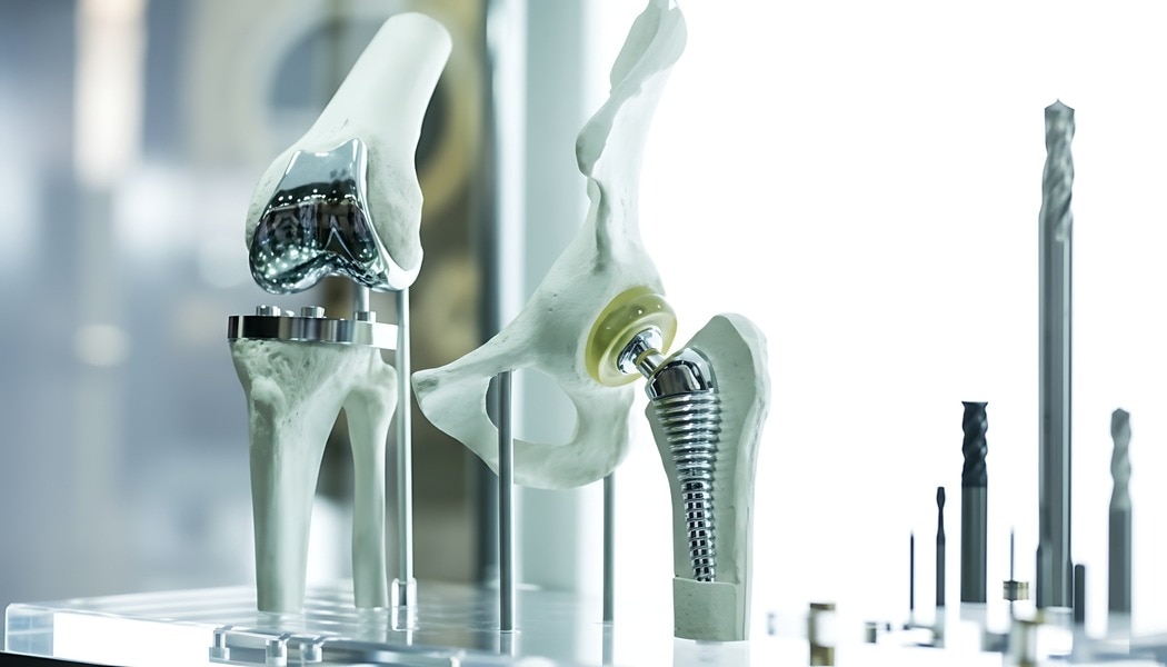
An uncemented revision stem gains fixation below damaged proximal bone, usually in the intact femoral diaphysis. Its porous or grit-blasted surface relies on a tight distal fit followed by bone ingrowth, unlike a primary femoral stem, which usually depends on the metaphysis and proximal canal.
| Option | Fixation strategy | Typical distinction |
|---|---|---|
| Primary femoral stem | Proximal press-fit and bone ingrowth | Shorter design for preserved metaphyseal bone |
| Standard uncemented stem | Proximal or metaphyseal press-fit | Not designed to bypass major osteolysis, fracture, or implant-removal defects |
| Uncemented revision stem | Distal axial and rotational press-fit | Longer, often tapered and fluted, to bridge proximal bone loss |
| Cemented revision stem | Cement fills the prepared canal | Provides fixation through cement rather than distal osseointegration |
Choose a revision stem when femoral loosening, osteolysis, an implant-removal defect, or a periprosthetic fracture leaves inadequate proximal support but preserves a usable distal fixation segment.
Paprosky type II and selected type III defects can suit distal fixation; severe type IV loss may lack enough diaphyseal bone and require impaction grafting, a custom implant, or proximal femoral replacement.
Do not select the longest stem automatically. Excessive length or diameter increases distal cortical contact, tip stress, thigh pain, stress shielding, and fracture risk. A straight stem can also strike the anterior cortex or perforate a bowed femur, so assess both anteroposterior and lateral images, with CT when the canal or deformity is unclear.
A modular revision stem restores version, offset, and leg length independently; a monoblock avoids a junction but sacrifices that control. Persistent infection still requires infection treatment, spacer planning, antibiotics, and viable host bone; mechanical fixation cannot compensate for unresolved periprosthetic joint infection.
How do you prove that the femur can support distal fixation?
Reliable distal fixation requires an intact, circumferential diaphyseal tube long enough to grip the femoral stem below the bone defect. Radiographs provide the first test: inspect full-length anteroposterior and lateral views for continuous cortices, measurable cortical thickness, canal diameter, and a straight fixation segment without fracture lines, cortical windows, osteolysis, or severe bowing.
| Finding | Supports fixation | Raises concern |
|---|---|---|
| Intact diaphysis | Continuous cortical contact for axial support | Short or interrupted fixation segment |
| Canal cross-section | Diameter matches the planned uncemented stem | Wide, irregular, or oval canal that prevents scratch fit |
| Cortical shell | Thick cortex resists stem settling and rotation | Thin cortex, cavitation, or segmental loss |
| Femoral bow | Stem follows the native canal on AP and lateral views | Straight stem contacts the anterior cortex or risks perforation |
| CT defect pattern | Circumferential bone remains around the fixation zone | Unseen posterior, medial, or lateral cortical deficiency |
CT is essential when radiographs hide cavitary or segmental loss. Review axial slices through the proposed fixation zone for a continuous cortical ring, the narrowest canal diameter, cortical thickness, and defects that could permit rotational toggling. CT also reveals cement or membrane that reduces usable length.
Do not treat a Paprosky label as proof. Confirm the canal after removing the loose implant, cement, membrane, and obstructing bone. If no adequate diaphyseal segment remains, a longer uncemented revision stem only shifts the problem; it does not create reliable fixation. Progressive postoperative subsidence confirms that axial or rotational support was inadequate.
How should stem geometry be planned without creating a new mechanical problem?
A stem that fixes securely in the distal femur can still place the hip in the wrong position.
Plan the fixation segment on calibrated AP and lateral radiographs, then use CT to confirm canal diameter, cortical thickness, femoral bow, and the length of intact diaphysis; a straight revision stem that fits the AP canal can strike the anterior cortex on the lateral view, perforate the femur, or create a high bending force.
- Select stem length and diameter for circumferential distal contact in the healthiest available diaphysis, not for maximum canal fill. Stop the stem above a severe bow or defect rather than forcing a long implant through a mismatched canal.
- Choose the fixation zone below osteolysis, fracture, or deficient cortex. Confirm that the remaining segment provides axial and rotational control; severe Paprosky Type IV loss may require another reconstruction rather than an uncemented stem.
- Size the proximal body to restore metaphyseal support without relying on it to rescue inadequate distal fixation. Account for an extended trochanteric osteotomy, because its distal limit and fixation must not undermine the planned fixation zone.
- Set offset, version, and leg length independently when anatomy demands it. A modular revision stem offers this control, while a monoblock uncemented stem avoids a junction but couples these variables.
- Use modular neck, body, and head options to restore soft-tissue tension and avoid impingement. Check the result for instability, excessive offset, reduced abductor leverage, and junction loading; large offsets and poor distal fixation raise fretting, corrosion, fracture, and disengagement risks.
Compare the planned hip implants in three dimensions, including the femoral bow and the acetabular component’s position. Choose head or neck length only after stem height, offset, and version are set, then verify leg length and range of motion on templated images.
Which mechanical, biological, and extraction problems must the reconstruction plan anticipate?
Plan for an uncemented revision stem around failure, not just fixation. Poor scratch fit results from undersizing, inadequate distal reaming, an irregular canal, thin cortices, or a bowed femur that contacts the anterior cortex before the stem reaches its fixation zone.
A straight stem in a pronounced bow can perforate the femur or create a high bending force.
| Problem | Main cause | Consequence |
|---|---|---|
| Poor axial or rotational fixation | Short intact diaphysis, undersizing, weak bone, inadequate reaming, or severe Paprosky Type IV loss | Subsidence, rotational instability, leg-length loss, and dislocation |
| Fracture or perforation | Hoop stress in thin cortices, eccentric reaming, forceful insertion, or canal-stem mismatch | Intraoperative fracture, cortical breach, and loss of the planned fixation zone |
| Failed biological fixation | Excessive micromotion, infection, or an interposed membrane between bone and implant | No osseointegration despite an initially tight radiographic fit |
Some initial settling is acceptable with a tapered fluted stem; progressive subsidence signals inadequate axial fixation, undersizing, poor bone quality, or rotational failure. An oversized or excessively distal stem can also cause stress shielding by carrying load away from the proximal femur, leaving that bone weaker.
Removal adds a separate hazard. A well-fixed femoral stem can strip cortical bone and require an extended trochanteric osteotomy, followed by cerclage fixation. Prepare extraction instruments, osteotomy equipment, and a backup stem; forcing removal can create additional bone loss and turn a salvageable reconstruction into a femoral replacement problem.
How can follow-up distinguish a stable stem from progressive failure?
A stable uncemented stem keeps the same height, alignment, and rotation on serial radiographs; progressive failure changes one of those relationships. Use matched AP pelvis and lateral femur images, then compare them with immediate postoperative films.
| Finding | Interpretation | Response |
|---|---|---|
| Unchanged stem position with developing spot welds or calcar remodeling | Bone ingrowth is progressing | Continue scheduled surveillance |
| Early settling that stops and remains aligned | Controlled seating | Observe with repeat imaging |
| Continued subsidence, varus tilt, widening radiolucent lines, pedestal formation, or a new fracture | Loss of fixation or failed ingrowth | Investigate promptly; revision may be necessary |
| New migration with pain, fever, drainage, or rising inflammatory markers | Mechanical failure, infection, or both | Obtain ESR, CRP, and hip aspiration before choosing reconstruction |
A well-fixed distal revision stem does not repair a deficient calcar, trochanteric nonunion, or abductor attachment. Persistent limp or recurrent dislocation therefore demands proximal reconstruction, trochanteric fixation, and restored offset—not simply a larger stem.
Control periprosthetic joint infection before definitive fixation. A porous implant cannot compensate for infected tissue; use staged treatment, an antibiotic spacer, and viable host bone when indicated.
Choose modularity only when independent adjustment of version, offset, height, or leg length outweighs taper-junction risks such as fretting, corrosion, fracture, or disengagement. A monoblock revision stem removes that junction but limits adjustment.
For sourcing hip implants, ask Komal Health Care Pvt Ltd for calcar-stem dimensions, compatible components, lot traceability, and surgical instrumentation; never substitute a cemented or bipolar component for a revision stem without system compatibility and fixation review.
Frequently asked questions
When is an uncemented revision stem appropriate?
Use it when damaged proximal bone cannot support fixation but the distal femoral diaphysis offers sufficient length, diameter, and cortical integrity for a tight fit and bone ingrowth.
How do you prove the femur can support distal fixation?
Review calibrated radiographs and CT scans for intact cortical bone, adequate diaphyseal length, canal shape, femoral defects, cortical perforation, and fracture risk.
What geometry problems must revision stem planning prevent?
Plan stem length, diameter, bow, taper, version, and offset together so the implant achieves distal fixation without malalignment, leg-length error, instability, or cortical contact.
Which complications should the reconstruction plan anticipate?
Anticipate intraoperative fracture, perforation, subsidence, loosening, stress shielding, thigh pain, infection, dislocation, and difficult future stem extraction.
How can follow-up show whether the stem is stable?
Compare serial radiographs for early migration, progressive subsidence, new lucent lines, pedestal formation, cortical changes, fracture, and loss of fixation.