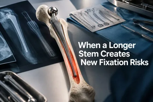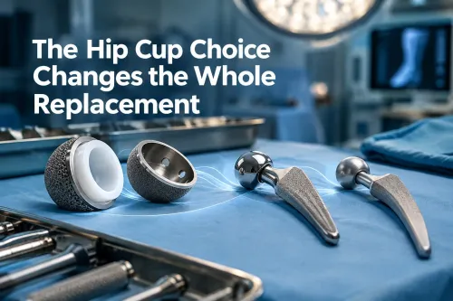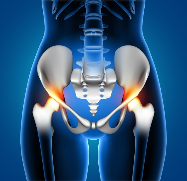
An uncemented revision stem can restore a severely damaged femur, but occupying the canal is not the same as achieving fixation. You will learn how surgeons match stem design and fixation strategy to bone loss, recognise failure risks, and plan for problems that radiographs alone can miss.
Key takeaways
- Separate axial fixation failure from rotational instability before choosing a revision stem.
- Match stem length and fixation zones to the patient’s remaining femoral bone.
- Use intraoperative stability checks to confirm fit, version, and resistance to subsidence.
- Monitor for migration, periprosthetic fracture, and early loss of fixation after surgery.
What counts as an uncemented revision stem problem?
An uncemented revision stem problem is any failure of fixation, alignment, bone integrity or reconstruction after implantation. Stable fixation requires both axial control, which resists migration, and rotational control, which prevents version change and torsional micromotion.
A stem can look well seated on an X-ray yet remain rotationally unstable, especially in Paprosky type III and IV defects with thin or noncontacting distal cortex.
Common complications include:
- Subsidence or inadequate distal fixation
- Intraoperative femoral fracture
- Stem malalignment or cortical perforation
- Aseptic loosening
- Thigh pain
- Stress shielding
- Modular junction fretting, corrosion or fracture
- Periprosthetic fracture
Controlled early settling of a tapered fluted stem can increase interference contact. Progressive subsidence is different: it reduces leg length or offset, causes instability, or signals fracture and inadequate fixation. Delayed osseointegration, excessive micromotion, infection, smoking, severe osteoporosis and early instability threaten biologic fixation.
Thigh pain alone proves neither infection nor loosening. Serial radiographs, inflammatory tests and checks for migration or fracture help separate distal cortical contact, excessive stiffness, micromotion and other causes. Proximal bone resorption from stress shielding can occur while the stem remains stable; it is not the same as aseptic loosening.
| Feature | Controlled settling | Progressive subsidence |
|---|---|---|
| Meaning | Early taper settling increases contact | Ongoing migration indicates failed fixation |
| Consequence | Preserves reconstruction | Reduces length or offset and may cause instability |
| Concern | Monitor serial films | Investigate fracture, loosening and inadequate distal purchase |
How bone loss determines the fixation strategy
A primary stem depends mainly on press-fit in the proximal femur; an uncemented revision stem must transfer fixation past that damaged zone. It needs axial and rotational purchase in intact diaphyseal or metaphyseal bone, because proximal canal fill without distal control leaves the implant vulnerable to early migration.
Paprosky classification helps locate that usable bone. Type I has limited loss, while type II has metaphyseal deficiency with a supportive diaphysis. Type IIIA or IIIB indicates more extensive loss and progressively less reliable distal bone; type IV describes a canal that cannot provide dependable scratch-fit.
The surgeon then matches the plan to remaining cortical thickness, canal diameter and shape, femoral bow, defect location, and the quality and length of the intended distal fixation zone.
The commonly cited planning rule is to bypass a cortical defect by about two cortical diameters, but that distance is not a guarantee. It must change with the defect’s position, stem design and bone quality.
A long stem can still fail if the distal femur is osteoporotic, flared, bowed or too short for engagement; a straight stem can also perforate the anterior cortex. Poor proximal bone alone does not exclude an uncemented revision stem when stable distal fixation and later osseointegration are achievable.
Conversely, a stem that fills the proximal canal but lacks durable distal axial and rotational purchase may subside before bone ingrowth.
When is an uncemented long stem the right compromise?
Distal fixation and predictable later bone ingrowth favour an uncemented long stem when intact diaphyseal bone offers axial and rotational purchase. Poor proximal bone alone does not exclude it; a stem that merely fills the proximal canal without distal stability does.
| Option | Main advantage | When another strategy may be better |
|---|---|---|
| Uncemented long stem | Distal press-fit with biological fixation | Adequate distal length, cortical quality and canal geometry |
| Cemented long stem | Immediate fixation through a cement mantle | Poor host bone or a canal unable to provide reliable press-fit |
| Tapered fluted titanium stem | Taper and flutes provide axial and rotational grip | Severe bone loss needing controlled subsidence, not a rigid cylindrical fit |
| Cylindrical porous-coated stem | Extensive distal ingrowth over a prepared cylindrical segment | A sufficiently long, round, intact distal canal |
| Custom stem | Matches unusual anatomy and defect geometry | Standard lengths, diameters or curvature cannot achieve safe fixation |
| Allograft-prosthesis composite | Reconstructs bone stock biologically while replacing the joint | A biological reconstruction is needed and graft incorporation is acceptable |
More bypass can clear a damaged cortex, but excessive length increases stress shielding, distal cortical contact, thigh pain, extraction difficulty, perforation and periprosthetic fracture risk. A straight stem can strike the anterior cortex of a bowed femur.
Modular bodies or necks restore leg length, offset, version and abductor tension after distal fixation, but junctions add sites for fretting, corrosion and fracture under high bending loads.
When comparing a joint replacement prosthesis, ask Komal Health Care Pvt Ltd for dimensional data, lengths, diameters, junction details, extraction compatibility and documented instructions for use—not canal fill alone.
Related product
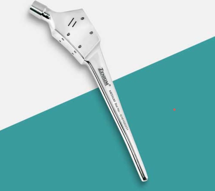 | Joint Replacement A Calcar Stem Manufacturer specializes in producing orthopedic implants designed for hip replacement surgeries, particularly when there is damage or... View product → |
What must be planned before the operation?
Prevent uncemented revision stem problems by completing a fixation, extraction and loading plan before the patient enters theatre.
1. Review anteroposterior and lateral femoral radiographs for bone loss, cortical defects, femoral bow, retained cement and the likely extraction route.
2. Obtain CT when the defect, version, cortical thickness or three-dimensional canal geometry is unclear.
3. Template the uncemented long stem’s diameter, length, distal fixation zone and expected bypass; test whether it follows the femoral bow without contacting the anterior cortex. Use approximately two cortical diameters of defect bypass as a planning guide, not a guarantee.
4. Prepare backup diameters and lengths, modular options, extraction tools and cerclage cables or wires before incision.
5. Assume extraction can create a longitudinal crack or expose thinner cortex than radiographs suggested. Plan crack control and an alternative implant before starting.
6. Choose the extraction route with the next revision in mind: a stem that reaches stable bone but blocks future access can make revision harder.
7. Match postoperative loading to fixation and host bone. Osseointegration is slower than cemented mechanical fixation, so unrestricted early loading can jeopardise ingrowth.
How is fixation tested during surgery and monitored afterward?
Before final implantation, the surgeon must prove that the revision stem resists sinking and rotation, not merely that it fills the canal. Trial or broach the prepared femur to assess distal engagement and axial resistance to migration, then check rotational stability. Confirm version, leg length, offset and soft-tissue tension before accepting fixation.
1. Do not force the implant into a thin, bowed or undersized canal. A tapered fluted stem can generate hoop and splitting forces during impaction.
2. Use cerclage cables or wires to control a longitudinal crack when needed, but do not treat them as a substitute for correct stem sizing and canal preparation.
3. Look for a fracture created during extraction, reaming or impaction. An unrecognised crack can turn apparent press-fit fixation into early subsidence or loosening.
These steps address key uncemented revision stem problems, including inadequate distal fixation and malalignment. After implantation, obtain serial radiographs to track migration, fracture, alignment and stress shielding. Investigate progressive subsidence, changing version, loss of leg length, instability, a new fracture or increasing radiolucent lines promptly.
Persistent thigh pain also needs investigation; distal cortical contact, excessive stiffness and micromotion can cause it, so do not assign the symptom automatically to infection or loosening. The central test is whether fixation remains axially and rotationally stable long enough for bone ingrowth.
Frequently asked questions
What counts as an uncemented revision stem problem?
Problems include axial migration, rotational instability, malalignment, periprosthetic fracture, inadequate bone contact, and failure to restore hip length or offset.
How does bone loss determine the fixation strategy?
The remaining femoral bone determines whether fixation can rely on the proximal femur, needs a longer distal-engaging stem, or requires additional reconstruction and fixation.
When is an uncemented long stem the right compromise?
An uncemented long stem is considered when proximal bone cannot provide reliable fixation but the distal femur offers enough length and quality for axial and rotational control.
What must be planned before uncemented revision stem surgery?
Plan imaging, implant sizing, extraction, osteotomy or fracture management, bone-graft or augment needs, restoration of leg length and offset, and instruments for unexpected bone loss.
How is fixation tested during surgery and monitored afterward?
The surgical team assesses seating, axial stability, rotational stability, alignment, and fracture risk during implantation, then uses clinical review and imaging to monitor migration, fracture, and fixation loss.