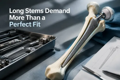
A long uncemented femoral stem can bypass missing proximal bone, but it also transfers fixation demands to the diaphysis, where poor sizing, femoral bow mismatch, or weak cortical bone can cause fracture, perforation, subsidence, or failed osseointegration. By the end, you will know which findings make this strategy reasonable, which warning signs require another reconstruction method, and how to assess fixation over time.
Key takeaways
- Use distal fixation when proximal femoral bone cannot support metaphyseal loading.
- Match stem diameter and geometry to the femur’s narrowest safe canal.
- Check for cortical contact, subsidence and migration during postoperative monitoring.
- Consider custom, modular, tapered or cemented reconstruction when a long stem is unsafe.
When does a difficult femur require more than a standard uncemented stem?
A standard uncemented stem is inadequate when the proximal femur cannot provide reliable fixation or its geometry makes metaphyseal loading unsafe. Revision surgery, Paprosky type IIIB or IV bone loss, an extended trochanteric osteotomy, a malunited fracture, severe osteoporosis, and proximal femoral deformity can shift fixation toward intact distal diaphyseal bone.
An uncemented long stem is not simply a longer version of a primary femoral stem: it depends on adequate distal length, cortical contact, and rotational control while the proximal femur contributes little initial support.
Before choosing an uncemented stem, measure or review:
- Anteroposterior and lateral hip radiographs for bone loss, canal shape, fractures, and implant position.
- Full-length femur images to identify anterior bowing; a straight stem can strike the anterior cortex, causing perforation, fissure, or a stress riser at its tip.
- CT when rotational anatomy, deformity, healed fracture malalignment, or femoral anteversion is unclear.
- The available intact distal diaphyseal length, canal diameter, cortical thickness, and bow radius.
- Offset, leg length, version, and the likely effect of distal fixation on proximal component orientation.
| Fixation choice | What changes the strategy | Main limitation |
|---|---|---|
| Standard uncemented stem | Adequate metaphyseal bone and normal geometry | Unreliable with major proximal deficiency |
| Uncemented long stem | Usable intact distal diaphysis with compatible bow | Perforation, fracture, subsidence, or poor rotation |
| Cemented long stem | Poor press-fit bone quality or inadequate distal grip | Cement mantle and infection concerns |
| Modular revision stem | Severe proximal loss requiring independent distal fixation and version adjustment | Junctional and rotational complications |
How do sizing and femoral geometry cause fractures or perforation?
The main uncemented long stem problems begin intraoperatively with poor fit: undersizing leaves inadequate diaphyseal contact, causing axial subsidence and weak rotational control, while oversizing generates excessive hoop stress. In thin osteoporotic cortex, forcing an oversized femoral stem into position can split the femur before fixation is secure.
Distal reaming can remove the cortex needed for distal fixation. An eccentric reamer may create a cortical defect; passing the femoral stem across that defect can extend it into a longitudinal fracture or redirect the tip through the cortex.
Risk rises sharply when the planned fixation crosses compromised bone:
- A previous osteotomy or healed fracture with malalignment
- Paprosky type IIIB or IV bone loss
- Severe osteoporosis or a large cortical defect
A straight uncemented long stem must match the femur’s shape, not only its canal diameter. In a bowed or deformed femur, the stem can strike the anterior cortex distally, causing perforation, an incomplete fissure, or a stress riser at the tip.
If intact diaphyseal bone is too short or narrow, increasing length only moves the fixation problem farther distally.
Review full-length anteroposterior and lateral femur images before reaming; standard hip films can hide the bow. Use CT when rotational anatomy or deformity is unclear, and confirm the available intact distal canal length and diameter. Stop if contact is eccentric, a defect lies in the fixation zone, or insertion requires force rather than controlled seating.
How can a stem look well positioned yet lose fixation after surgery?
An uncemented long stem can look well positioned on the first postoperative film yet lack durable fixation. Primary mechanical stability comes from press-fit contact in intact diaphyseal bone; osseointegration comes later. Excessive micromotion, poor bone quality, inadequate distal engagement, or poor rotational control can prevent bone ingrowth.
Early axial subsidence may be acceptable when it is small and stops, but progressive migration can shorten the leg, change offset, destabilise the hip, and eliminate distal fixation.
Serial radiographs separate settling from failure:
- Stable settling: minor early subsidence, preserved stem alignment, no expanding radiolucency, and no new fracture line.
- Concerning migration: continuing subsidence, changing varus or valgus alignment, or a new radiolucent line around the distal fixation zone.
- Distal overload: pedestal formation or cortical remodeling at the stem tip, indicating concentrated load transfer.
- Mechanical failure: fracture lines, progressive radiolucency, or loss of contact with the diaphyseal cortex.
A stiff distally engaging femoral stem can cause thigh pain from distal cortical contact or stress concentration even when it remains fixed. Do not label that pain benign until radiographs exclude subsidence, loosening, fracture, or perforation.
Distal fixation also unloads the proximal femur, producing stress shielding and bone resorption that can complicate later revision, even if the stem remains stable. This fixation trade-off matters when comparing hip implants, because a longer stem is not automatically a more secure one.
When is another reconstruction strategy safer than a long uncemented stem?
Choose another reconstruction when the femur cannot provide reliable distal cortical contact for an uncemented long stem. A longer implant does not create missing bone; it can shift fixation into a bowed, thin, or perforation-prone segment. These uncemented long stem problems include fracture, inadequate rotational control, subsidence, and later stress shielding.
| Option | Fixation logic | When it fits | Main limitation |
|---|---|---|---|
| Cemented long stem | Cement fills the canal and transfers load along the prepared cement mantle. | Poor-quality bone or a canal that cannot deliver a dependable press fit, provided infection is controlled. | A distorted canal can produce an uneven mantle or perforation; cement pressurisation can fracture thin cortices, and it does not treat active infection. |
| Calcar-replacing stem | Replaces deficient proximal medial support and restores neck height and offset. | Proximal bone loss with enough remaining geometry for stable fixation, often when distal diaphyseal engagement is not the central defect. | It is unsafe when the proximal femur cannot support the implant or when deformity prevents accurate seating. |
| Modular tapered fluted stem | Flutes obtain distal axial and rotational fixation while a separate proximal body restores length and offset. | Paprosky IIIB or IV loss, revision surgery, or an extended trochanteric osteotomy with adequate intact diaphyseal length. | The junction adds fretting, corrosion, fracture, and rotational-loss risks; poor distal bone still defeats fixation. |
| Allograft-prosthesis composite | Reconstructs missing bone while the prosthesis replaces the joint. | Large structural defects and selected younger patients when biological reconstruction is realistic. | Union, graft resorption, fracture, and infection remain serious concerns, making it unattractive after active or recently treated infection. |
If intact diaphyseal fixation is short, osteoporotic, sharply bowed, or interrupted by deformity, do not solve the problem by choosing a longer uncemented stem. CT and full-length imaging should confirm a safe fixation segment before selecting hip implants.
What should you verify before choosing and monitoring the implant?
Before selecting an uncemented long stem, review full-length anteroposterior and lateral femur radiographs, not only hip views. Use CT when rotational anatomy, a healed fracture, or deformity makes the canal path uncertain; measure intact distal diaphyseal length, canal diameter, femoral bow, and the planned stem tip position.
- Confirm that the femoral stem matches the anterior bow and does not force distal cortical contact.
- Define the minimum circumferential cortical engagement needed for axial and rotational stability.
- Mark defects, osteotomy lines, fracture risk, and the available zone for cerclage cables, plates, or cortical strut reinforcement.
- Compare the stem’s taper, diameter increments, length, offset, material, coating, and distal fixation zone with the femur measurements.
- Record the implant lot, UDI, surgical technique, contraindications, and radiographic landmarks before implantation.
| Evidence | Compare | Why it matters |
|---|---|---|
| Full-length radiographs | Bow, defects, and stem-tip clearance | Detects perforation or a stress riser hidden on hip films |
| CT and canal measurements | Rotational anatomy and intact diaphyseal length | Exposes inadequate distal fixation |
| Implant documents | Drawings, sizes, taper, alloy, coating, and IFU | Prevents mismatched components or unplanned sizing |
When comparing hip implants from Komal Health Care Pvt Ltd with alternatives, place these documents side by side rather than relying on catalogue names.
Use the immediate postoperative full-femur image as a baseline, then compare scheduled serial radiographs. Stable settling differs from progressive migration: investigate increasing subsidence, changing alignment, distal radiolucency, pedestal formation, cortical remodeling at the tip, or a fracture line. A single early image cannot prove durable fixation.
Frequently asked questions
When does a difficult femur require more than a standard uncemented stem?
Consider a longer stem when revision surgery, Paprosky type IIIB or IV bone loss, an extended trochanteric osteotomy, malunited fracture, severe osteoporosis or proximal femoral deformity prevents reliable proximal fixation.
How do sizing and femoral geometry cause fractures or perforation?
An oversized stem can split weak cortical bone, while mismatch between stem shape and a bowed, narrow or deformed femur can drive the tip into the cortex or perforate the canal.
How can a stem look well positioned yet lose fixation after surgery?
Radiographs can show acceptable alignment while inadequate distal contact, poor axial stability, subsidence, migration or progressive radiolucency reveals loss of fixation during follow-up.
When is another reconstruction strategy safer than a long uncemented stem?
Choose another strategy when the femur cannot provide a continuous, safe distal fixation segment or when its deformity makes stem passage likely to cause fracture or cortical perforation.
What should you verify before choosing and monitoring the implant?
Verify the femoral defect pattern, canal diameter and bow, available distal fixation length, stem geometry, intraoperative stability and serial radiographs for subsidence, migration, fracture and loosening.