A screw can loosen, migrate, bend, break, strip, or enter a joint for very different reasons, and the same radiographic finding does not always indicate the same failure mechanism. By the end, you will be able to distinguish mechanical failure from infection or planned dynamization, recognise urgent warning signs, and understand how clinicians investigate a suspected fixation problem.
Key takeaways
- Classify screw failure as loosening, migration, breakage, pullout, or malposition.
- Compare screw changes with fracture healing before judging fixation failure.
- Seek urgent assessment for worsening pain, deformity, drainage, fever, or new numbness.
- Prevent failure by matching screw design and fixation strategy to bone quality.
Classify the screw problem before judging the fixation
A screw can fail by losing purchase, pulling out, bending, breaking, migrating, backing out, stripping its threads, or sitting in the wrong position. These orthopedic implant screw problems are not interchangeable: a loose screw from poor bone purchase differs from a broken screw beside a nonunion, and intentional dynamization differs from uncontrolled back-out.
| Screw design | Characteristic failure pattern | What to check |
|---|---|---|
| Cortical | Stripping, breakage, or loss of purchase in thin cortex | Thread engagement, pilot-hole size, trajectory, and torque |
| Cancellous | Pull-out or loosening after over-tightening or cyclic loading | Bone quality and whether insertion torque stripped trabecular purchase |
| Cannulated | Guidewire bending, migration, breakage, or joint entry; the screw may follow that error | Guidewire position in two fluoroscopic projections before drilling |
| Locking | Hole-adjacent fatigue, screw breakage, or delayed union in an overly rigid construct | Working length, plate stiffness, screw distribution, and fracture biology |
| Lag or malleolar | Loss of compression, back-out, tendon irritation, or intra-articular penetration | Thread position, screw length, reduction, and joint clearance |
Length is functional, not cosmetic. A short screw may miss the far cortex or provide inadequate engagement; a long one can damage cartilage or abrade a tendon. Back-out or a radiographic halo does not prove infection: assess purchase, trajectory, reduction, loading, and union.
Fatigue breakage after repeated loading often signals nonunion or construct instability rather than a single overload event. These are central bone screw complications.
Trace failure to bone quality, technique, or construct mechanics
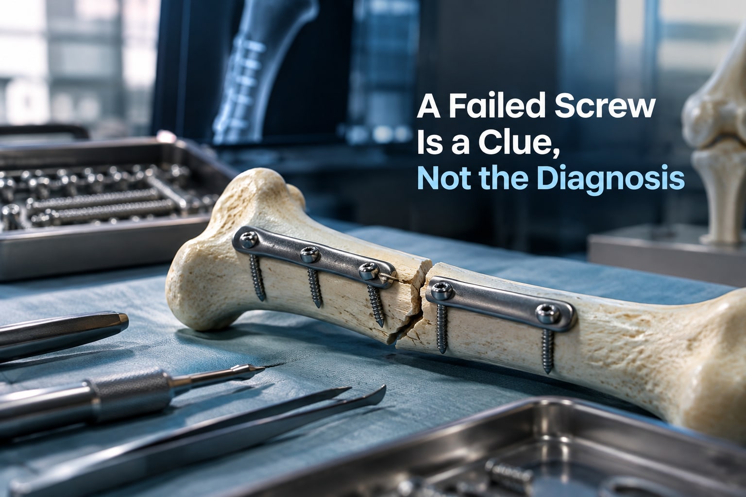
A screw can survive implantation and early weight bearing, then break after repeated loading when fracture union is delayed or absent. The broken screw often signals unresolved nonunion or construct instability, not a defective screw alone.
Trace the failure through three checkpoints:
- Bone quality: Osteoporotic or thin cancellous bone provides shallow thread purchase. Over-tightening strips trabeculae during insertion even when the head appears seated, so final seating torque does not prove fixation strength.
- Technique: An oversized hole, over-tapping, an unpreserved far cortex, or a pre-tapped hole reduces engagement. Cortical screw concerns also include omitting the glide hole needed for effective lag compression. A bent or misplaced guidewire can send a cannulated screw into the wrong trajectory or joint.
- Construct mechanics: A short working length, excessive plate stiffness, too few screws near the fracture, poor plate contour, wrong screw length, loss of reduction, or loading before union concentrates cyclic force. The result can be loosening, pull-out, bending, back-out, or fatigue breakage.
Removal becomes difficult when the head is stripped, the screw is cross-threaded, cold-welded to a locking plate, buried by bone, or itself bent or broken. A radiographic halo or migrating screw does not establish infection; assess purchase, alignment, union, and inflammatory or infectious evidence together.
These orthopedic screw fixation problems require different solutions because treating a loose screw without correcting nonunion or instability leaves the cause in place.
Recognise warning signs and know when assessment is urgent
Emergency assessment is needed after fracture fixation for sudden severe pain, a new deformity, or any sign that blood flow or nerve function has changed. Do not wait for a routine appointment if the limb becomes cold, pale, blue, numb, or weak, or if swelling rapidly increases.
Seek emergency care for:
- Pain that escalates despite prescribed medicine, especially with a tense swelling or pain when the fingers or toes are passively stretched; this can indicate compartment syndrome.
- New inability to move the hand, foot, fingers, or toes, or loss of sensation below the fixation.
- Sudden shortening, rotation, or visible deformity after a click, fall, or loading event.
- Heavy bleeding, wound opening, or metal becoming visible.
- Chest pain, sudden breathlessness, coughing blood, or fainting after surgery; these symptoms can signal a pulmonary embolism.
Arrange same-day medical assessment for persistent or worsening pain, redness spreading from the incision, increasing warmth or swelling, fever, chills, pus, or drainage that continues beyond expected wound healing. These orthopedic implant issues can reflect infection, loosening, loss of reduction, or nonunion, not merely normal recovery.
| Finding | Action |
|---|---|
| New limb coldness, pallor, numbness, or weakness | Emergency department immediately |
| Rapid swelling with severe pain | Emergency assessment now |
| Drainage, fever, or progressive redness | Same-day surgical review |
| Gradually increasing load-related pain | Prompt orthopaedic assessment |
Understand how clinicians investigate suspected screw failure
A single radiograph cannot show whether an orthopedic implant screw problem comes from infection, nonunion, or mechanical overload. Clinicians compare immediate postoperative images with serial anteroposterior, lateral, and oblique views, then match changes with pain, drainage, fever, tenderness, and function. A suspected shift on one projection may be projectional rather than true migration.
Investigation usually combines:
- Infection: examine the wound and obtain a complete blood count, C-reactive protein, and erythrocyte sedimentation rate. Persistent drainage, rising markers, or unexplained loosening may prompt image-guided aspiration or operative deep-tissue and implant-associated cultures; superficial swabs are not a reliable substitute.
- Migration or loss of reduction: compare screw position, fracture alignment, plate relationship, and joint clearance across time. CT clarifies three-dimensional penetration, subtle displacement, and bone loss when plain films conflict with the examination.
- Breakage: inspect all views for a discontinuity, then use CT to locate fragments and determine whether the fracture has bridged. A broken screw after repeated loading often signals delayed union or nonunion rather than proving that the screw initiated failure.
- Nonunion: assess serial bridging callus, persistent fracture lines, motion or tenderness, and the biological context. CT can confirm an incomplete union, while infection testing remains important because infected nonunion can resemble mechanical failure.
Clinicians therefore interpret imaging as a timeline, not a verdict. They assess the whole construct, loading history, bone quality, and insertion records before deciding whether observation, protected weight bearing, further sampling, or revision is appropriate.
Reduce preventable failure through system selection and fixation planning
Final seating torque does not prove fixation strength. In cancellous or osteoporotic bone, over-tightening can strip trabecular purchase while the screw head still looks correctly seated. Use a torque-limiting driver and record the insertion method rather than treating driver resistance as a strength test.
Match the screw, hole preparation and construct to the bone and fracture. A cortical screw used for lag compression needs a glide hole in the near fragment; without it, the threads engage both fragments and produce little controlled compression.
Preserve the far cortex, use the specified drill and tap sizes, and do not substitute an oversized hole or over-tap.
Osteoporotic fixation benefits from a planned load path, not simply a larger screw or higher torque. Consider longer plates, distributed fixation, safe bicortical purchase, fixed-angle interfaces and augmentation when indicated; balance each against soft-tissue exposure, joint penetration and cement leakage.
Plan for cyclic loading because delayed union can break a screw after weeks of apparently successful use.
Before closing, document decisions that allow later investigation of bone screw complications and orthopedic screw fixation problems:
- Implant type, diameter, length, material and lot number
- Drill, tap and glide-hole sizes
- Insertion torque setting and whether purchase was satisfactory
- Cortical engagement, plate contour, screw distribution and final imaging
- Weight-bearing restrictions and the biological or mechanical reason for them
When reviewing components from Komal Health Care Pvt Ltd, request this traceability alongside the device specifications so a later failure can be assessed against the intended construct, not guessed from the broken screw alone.
Related products
 | Bone Nails ₹550 Our range of interlocking nail, intramedullary nail, fracture nail are can be used in 35 different cases of complex fractures. View product → |
 | Orthopaedic ₹1200 We offer orthopedic implants, orthopedic surgical implants as bone screws for orthopedic usage. View product → |
Frequently asked questions
What are the main types of orthopedic implant screw problems?
Problems include loss of purchase, pullout, bending, breakage, migration, backing out, stripped threads, and incorrect screw position. Each indicates a different mechanical or clinical issue.
What causes orthopedic screw fixation problems?
Common causes include poor bone quality, inadequate purchase, incorrect drilling or insertion, excessive loading, an unstable fracture pattern, and nonunion that keeps stress on the construct.
When does a suspected screw problem require urgent assessment?
Seek urgent assessment for severe or increasing pain, new deformity, wound drainage, fever, sudden loss of function, new numbness, colour change, or a screw that appears to threaten skin or a joint.
How do clinicians investigate suspected screw failure?
They review symptoms, the operation and implant records, examine the limb and compare serial X-rays. CT can define fracture healing, screw position, loosening, and joint involvement when plain films are unclear.
How can preventable orthopedic implant screw problems be reduced?
Clinicians reduce risk by assessing bone quality, choosing an appropriate screw and plate or nail system, obtaining adequate purchase, avoiding malposition, and planning fixation around expected loads and fracture healing.
Related product
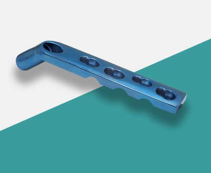 | Bone Plate Dhs Locking Plate Manufacturer DHS (Dynamic Hip Screw) locking plate manufacturers produce specialized orthopedic implants used in the surgical treatment of proximal femur... View product → |