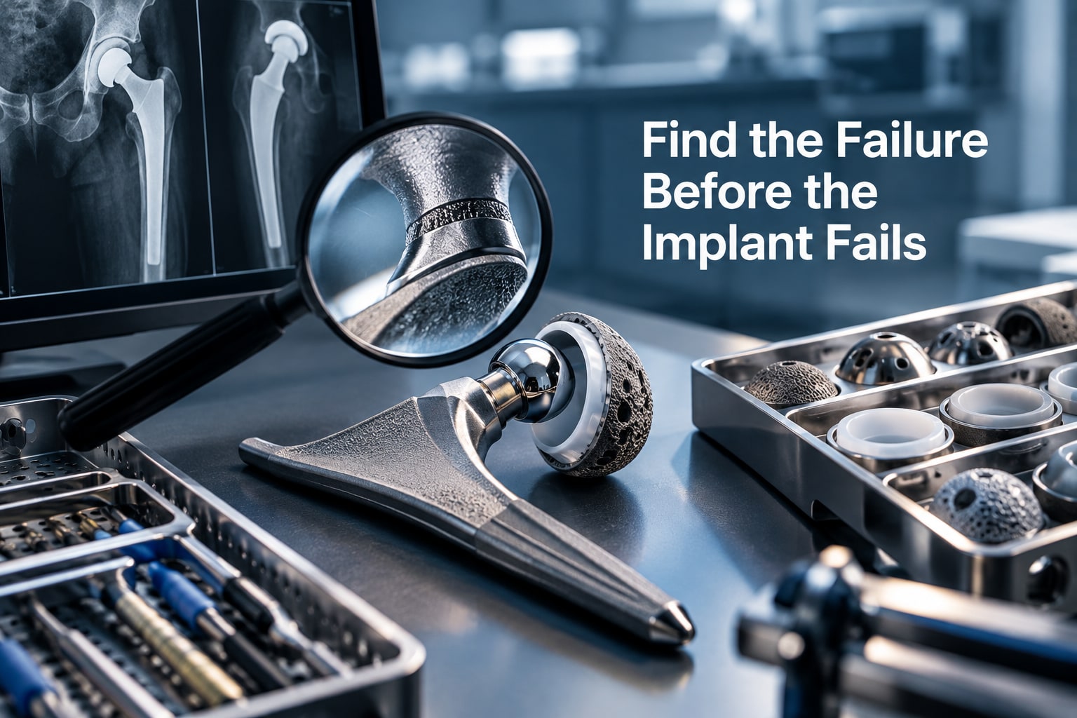
A painful or unstable hip replacement does not identify its own cause: loosening, infection, malposition, fracture, wear and soft-tissue reactions require different investigations. Hospitals can triage the warning signs, compare the right records and images, and determine when observation is unsafe, revision consultation is urgent, or emergency treatment is required.
Key takeaways
- Treat dislocation, infection, fracture, and nerve deficits as urgent problems.
- Compare serial radiographs for migration, bone loss, loosening, and fracture.
- Use targeted tests to distinguish infection, implant failure, and soft-tissue pain.
- Record batch details and confirm component compatibility before revision surgery.
Classify the problem before deciding how urgently to act
Treat total hip replacement implant problems by consequence, not by the label “failed hip.” A dislocation, suspected infection, periprosthetic fracture or new nerve deficit can threaten the limb or patient quickly; a painless radiographic change needs a different pathway.
| Problem pattern | Action | Trigger |
|---|---|---|
| Instability or dislocation | Emergency assessment | Sudden deformity, shortening, severe pain or inability to bear weight |
| Fracture or component breakage | Emergency assessment | Fall, acute thigh or groin pain, new shortening or inability to walk |
| Infection | Emergency assessment | Fever, wound drainage, rapidly worsening pain or sepsis signs |
| Nerve injury | Emergency assessment | New foot drop, numbness or marked weakness |
| Loosening, migration or progressive osteolysis | Urgent revision review | New weight-bearing pain, subsidence, radiolucent lines or bone loss |
| Corrosion or adverse local tissue reaction | Urgent revision review | New pain, swelling, weakness or metal-on-metal concern |
| Leg-length, offset or malposition problem | Urgent review | Limp, instability, impingement or persistent functional deficit |
| Wear without acute symptoms | Routine investigation | Progressive polyethylene wear, osteolysis or heterotopic ossification on surveillance images |
Routine does not mean ignore. Compare serial, correctly positioned radiographs and obtain the operative implant record; a single baseline image cannot show movement over time. Persistent pain or loosening also needs infection testing, including inflammatory markers and, when indicated, aspiration, because low-grade periprosthetic joint infection can lack fever or drainage. Sudden deterioration overrides routine scheduling.
Use the patient record and targeted tests to separate failure from other pain
A pain timeline often separates normal recovery from failure. Record the operation date, onset and location of pain, whether it is improving, and any injury, wound problem, fever, clicking, instability or sudden loss of weight-bearing.
Retrieve the operative report, implant stickers, prior radiographs and details of the joint replacement prosthesis, including component positions and bearing couple.
Examine gait, leg length, hip range of motion, abductor strength, Trendelenburg sign, trochanteric tenderness and the lumbar spine. Check sensation, reflexes and motor power for nerve or radicular disease. Groin pain with rotation points toward the hip; lateral tenderness suggests bursitis; buttock pain with back movement or a positive nerve-tension test supports spinal pain.
Weak abductors can explain limp without proving that the hip implants have failed.
Use targeted tests rather than ordering every scan:
- Obtain anteroposterior pelvis and lateral hip radiographs, comparing them with earlier images for migration, fracture, dislocation, lucent lines or wear.
- Order erythrocyte sedimentation rate and C-reactive protein when infection is possible; aspirate the joint for synovial white-cell count, differential, culture and, where indicated, additional validated infection tests.
- Use ultrasound for a trochanteric bursa or fluid collection, and metal-artifact-reduction MRI for adverse tissue reaction or muscle damage.
- Use calibrated imaging when true leg-length or offset change is suspected; subjective shortening is unreliable.
No fever or drainage does not exclude low-grade infection. Progressive pain, weakness, instability or deteriorating function deserves investigation rather than reassurance based on an apparently well-fixed implant.
Read serial radiographs for movement, bone loss and fracture
Compare every follow-up image with the immediate postoperative baseline and the most recent study, using the same AP pelvis and lateral hip views when possible. A single film shows position; serial films show change. Check image quality and pelvic rotation first, because poor positioning can mimic migration or leg-length discrepancy.
| Finding | AP pelvis comparison | Lateral hip comparison | Concern |
|---|---|---|---|
| Progressive radiolucency | Lines at the acetabular shell or femoral stem–bone interface; note widening or new zones | Lines around the stem’s anterior or posterior interface | Aseptic loosening |
| Migration or subsidence | Cup movement, altered inclination, changing hip centre, or stem sinking relative to fixed landmarks | Stem sinking, altered version, or loss of seating | Component movement |
| Wear and osteolysis | Eccentric femoral head position, superior migration, and expanding bone defects | Posterior or anterior osteolysis and head position | Polyethylene wear with bone loss |
| Fracture or breakage | New cortical break, screw failure, or altered component alignment | Fracture around the stem, displaced fragments, or a broken modular part | Periprosthetic fracture or implant failure |
| Heterotopic ossification or stress shielding | New bone around the hip or proximal femoral bone-density loss | Extent of new bone and its effect on motion | Functional restriction or altered load transfer |
Measure changes against fixed landmarks such as the teardrop, acetabular roof, lesser trochanter, and stem shoulder. Record migration in millimetres when the films are calibrated. Highly cross-linked polyethylene reduces wear but does not prevent it; progressive osteolysis can loosen THR implants before pain becomes severe.
A well-fixed image also does not exclude soft-tissue or bearing failure.
Investigate hidden soft-tissue, bearing and modular-junction failures
A normal-looking radiograph does not exclude important total hip replacement implant problems. Match the suspected failure to laboratory sampling and cross-sectional imaging rather than treating all hip implants as equivalent.
- Infection: measure C-reactive protein and erythrocyte sedimentation rate, then aspirate the joint for synovial white-cell count, neutrophil percentage, aerobic and anaerobic cultures, and alpha-defensin when indicated.
- Metal debris or adverse local tissue reaction: identify the bearing couple, measure blood cobalt and chromium, and use ultrasound or metal-artifact-reduction MRI to find fluid, pseudotumour, muscle damage or bone destruction. Blood levels support assessment; they do not prove or exclude tissue injury.
- Polyethylene wear: compare correctly positioned serial films, then use CT to define osteolysis, liner position and hidden bone loss when plain images are uncertain.
- Ceramic damage: investigate new squeaking, grinding or sudden loss of function with CT and cross-sectional imaging. Assess the ceramic head, liner, taper and retained fragments together.
- Corrosion: inspect the modular head-neck taper and other junctions for fretting or mechanically assisted crevice corrosion, using serum metals and metal-artifact-reduction MRI when symptoms or a mass warrant it.
- Component breakage: obtain thin-slice CT and compare operative records with prior images to locate a fractured stem, neck, head, liner or fixation element.
A ceramic fracture requires management of ceramic debris and taper damage, not just head exchange. Corrosion can occur with ceramic-on-polyethylene bearings, so a radiographically fixed hip does not end the investigation.
Protect patients with traceability and compatible component decisions
A joint replacement prosthesis is traceable only when every implanted part connects to one patient and one operative record. For each total hip replacement, retain the manufacturer, catalogue number, lot or serial identifier, implant stickers, side, size and operative date for every modular part.
| Component | Record to retain | Why it matters |
|---|---|---|
| Acetabular shell, liner and screws | Manufacturer, catalogue, lot or serial numbers | Identifies the complete socket assembly during a recall or revision |
| Femoral stem and head | Manufacturer, catalogue, lot or serial numbers; size and taper details | Confirms the original geometry and prevents an incompatible replacement head |
| Adapters or other modular parts | Part number, lot or serial number and instructions for use | Reveals combinations that were not authorised or tested together |
Before revision, compare the proposed components against the original manufacturer’s instructions for use. Matching diameter alone is not enough: taper geometry, locking mechanism, material pairing and documented regulatory authorisation determine compatibility. An incorrect combination can cause corrosion, wear, dissociation or poor fixation.
Ask suppliers to provide legible labels, complete lot records, approved component combinations, revision notices and a named recall contact. Hospitals should also record which patients received each lot, so a recall reaches people rather than stopping at the storeroom.
When hospitals assess suppliers such as Komal Health Care Pvt Ltd, they should test whether its documentation supports this patient-level traceability for THR implants, not merely whether the component looks dimensionally suitable.
Related products
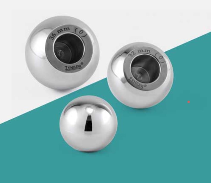 | Joint Replacement An Internal Head Manufacturer specializes in producing internal femoral heads used in hip replacement systems. View product → |
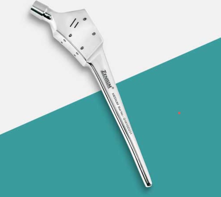 | Joint Replacement A Calcar Stem Manufacturer specializes in producing orthopedic implants designed for hip replacement surgeries, particularly when there is damage or... View product → |
Frequently asked questions
How should hospitals classify total hip replacement implant problems?
Classify them by consequence and urgency: dislocation, suspected infection, periprosthetic fracture, and new nerve deficit require rapid assessment, while painless radiographic changes follow a different pathway.
Which tests help separate implant failure from other causes of hip pain?
Review the patient record and use targeted tests to distinguish infection, mechanical failure, soft-tissue causes, bearing problems, and modular-junction failure.
What should clinicians look for on serial radiographs?
Compare images over time for component movement, progressive bone loss, radiolucent changes, migration, and periprosthetic fracture.
How can hospitals reduce risk during revision or component replacement?
Maintain implant traceability, record component and batch details, and confirm that replacement parts are compatible with the existing joint replacement prosthesis.
Related products
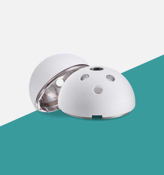 | Joint Replacement The Acetabular Cup is a key component in total hip replacement surgery, designed to fit into the hip socket (acetabulum). View product → |
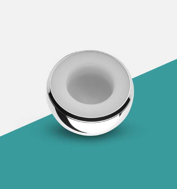 | Joint Replacement Modular Bipolar Cup Manufacturer A Modular Bipolar Cup Manufacturer specializes in producing advanced orthopedic components used in hip replacement surgeries. View product → |
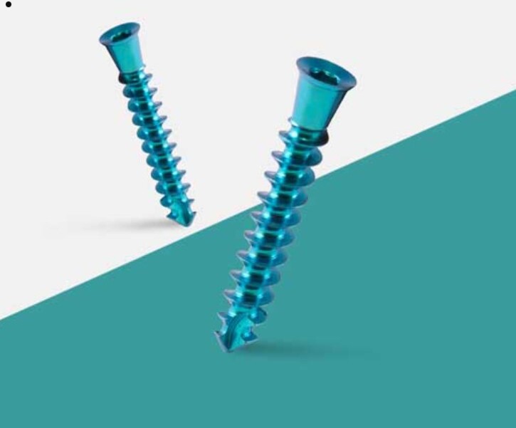 | Joint Replacement A THR Screw Manufacturer produces specialized screws used in Total Hip Replacement (THR) surgeries to secure implants and enhance joint stability. View product → |
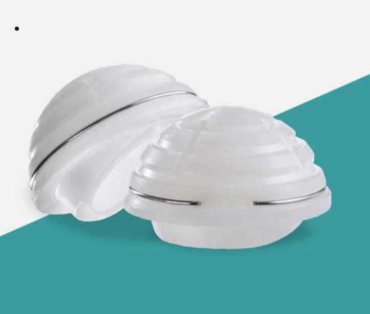 | Joint Replacement Acetabular Cup Cemented Manufacturer An Acetabular Cup Cemented Manufacturer produces acetabular components specifically designed for cemented fixation in total hip replacement... View product → |