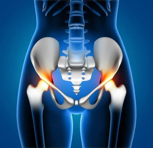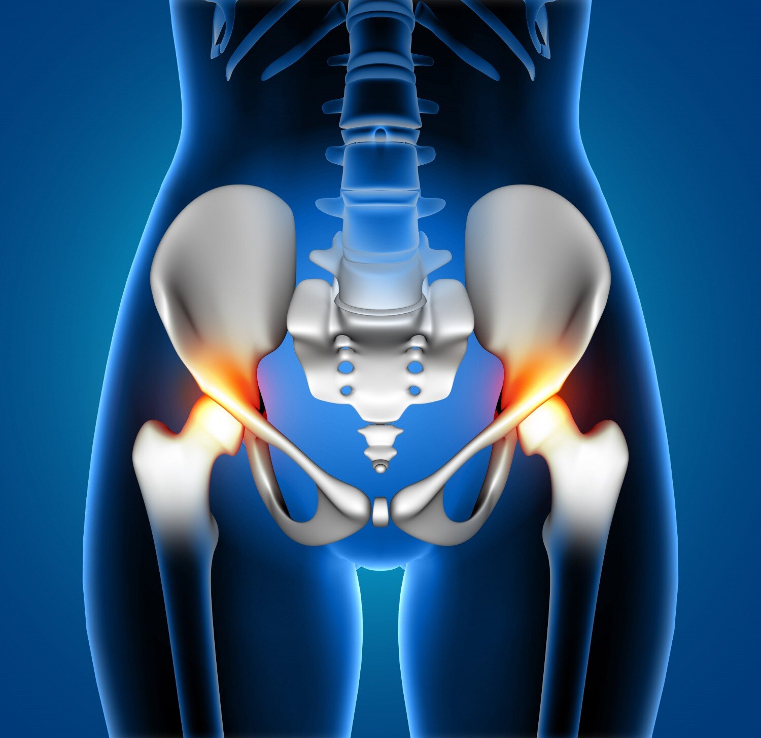
Pain after hip replacement does not identify the failed part by itself: infection, loosening, wear, fracture, instability and spine or tendon disorders can produce similar complaints. By checking symptom timing, serial radiographs, infection tests, implant records and bone loss in the right order, you can separate an urgent complication from a problem that needs planned revision assessment.
Key takeaways
- Match pain, migration and instability patterns to the likely complication.
- Compare serial radiographs before calling a cemented cup loose.
- Rule out infection and identify whether the cup, cement or bone is failing.
- Use the cement mantle and operative record to guide revision planning.
Match the symptom pattern to the likely complication
A cemented-cup problem is more than a loose cup. Cement–bone debonding, cement mantle fracture, cup migration or displacement, polyethylene wear, osteolysis, infection, instability, dislocation, leg-length change and persistent groin pain can all involve the cemented acetabular construct.
Timing changes the differential. Pain during the first postoperative days may reflect wound or soft-tissue injury, early infection, fracture, instability or an acute technical problem; new pain years later raises concern for loosening, wear, osteolysis, infection or late dislocation.
| Symptom pattern | More likely concern | What to do |
|---|---|---|
| Sudden inability to bear weight, shortened or rotated leg, new deformity, or severe pain after a fall | Dislocation or periprosthetic fracture | Seek urgent clinical assessment |
| Fever with worsening hip pain, wound drainage, warmth or rapidly increasing redness | Periprosthetic joint infection | Obtain urgent medical review |
| Progressive groin or thigh pain during weight bearing | Mechanical loosening, cement debonding or mantle fracture | Arrange orthopaedic assessment |
| Sudden pain with deformity and loss of motion | Dislocation or cup displacement | Seek urgent assessment |
| Gradual pain, reduced motion or increasing stiffness | Polyethylene wear with osteolysis | Arrange imaging and review |
| Groin pain when climbing stairs or flexing the hip | Iliopsoas irritation or impingement | Discuss examination with the treating clinician |
| Worsening function without fever or drainage | Low-grade infection or mechanical failure | Do not exclude infection |
Clicking alone does not prove cup failure. Lumbar-spine disease, trochanteric pain and other joint replacement prosthesis problems belong in the differential diagnosis of hip implant complications.
Use serial radiographs before deciding that the cup is loose
Confirm suspected cemented acetabular cup problems with examination and serial imaging, not one radiograph.
- Observe gait and weight-bearing.
- Measure leg length and hip range of motion.
- Test instability and assess the wound.
- Examine the spine and abdomen for referred pain.
- Palpate the greater trochanter for tenderness.
Compare current anteroposterior pelvis and cross-table lateral radiographs with the immediate postoperative films. Keep pelvic rotation and tilt consistent; positioning changes can mimic migration. Map radiolucency in the three DeLee–Charnley acetabular zones.
| Finding | Interpretation | Concern |
|---|---|---|
| Stable, narrow radiolucent line | May represent an established interface | Does not prove loosening |
| Progressive or complete radiolucency | Suggests interface failure | Greater concern, especially with migration |
| Superior or medial migration | Demonstrates component movement | Stronger evidence of mechanical failure |
| Changed inclination, mantle fracture or broken fixation feature | Indicates construct disruption | Supports loosening or displacement |
| Increasing head–cup eccentricity, osteolysis or fracture | Suggests wear or bone damage | Requires assessment even if the cup looks intact |
Migration is stronger evidence of mechanical failure than one unchanged radiolucent line. Look for polyethylene wear, osteolysis and periprosthetic fracture on every comparison.
Order CT when radiographs do not define retained cement, pelvic discontinuity, component position or acetabular bone loss. Metal artifact can obscure findings, so CT supports revision planning but does not replace C-reactive protein, erythrocyte sedimentation rate or hip aspiration when infection remains possible.
Rule out infection and identify which component is failing
Radiographs cannot reliably separate infection from aseptic loosening: both can produce radiolucent lines, migration and bone loss. Before revision for painful hip implants, review wound drainage, redness, fever and prior wound treatment, then obtain serum C-reactive protein and erythrocyte sedimentation rate when infection remains possible.
| Finding | Points toward | Next check |
|---|---|---|
| Progressive stem subsidence or fracture | Femoral-component failure | Compare serial femoral radiographs |
| Head damage or increasing head–cup eccentricity | Bearing or liner wear | Assess polyethylene loss and osteolysis |
| Osteolysis behind a stable cup | Wear-debris disease | Map bone defects; do not assume cup loosening |
| Pelvic or trochanteric tenderness | Bone or soft-tissue disease | Examine abductors, trochanter and pelvis |
| Instability, impingement or spinal pain | Non-cup pain source | Test stability, lumbar spine and gait |
If markers or the clinical picture warrant it, aspirate the hip for synovial white-cell count, differential and culture. Interpret thresholds against time from surgery, inflammatory arthritis and recent antibiotics; a normal CRP or ESR and one negative aspiration culture do not exclude low-grade periprosthetic joint infection.
If revision proceeds, collect at least three, preferably five or six, deep tissue specimens before antibiotics when clinically safe. Label each separately; pooled samples make the result difficult to interpret. These steps prevent cemented cup problems from being mistaken for femoral failure, infection, wear, adverse tissue reaction or referred pain.
Read the cement mantle and operative record for the failure mechanism
Cement quality means a complete, bonded mantle without voids, cracks or interface separation—not simply an undamaged cup. Inspect the cement–bone and cement–implant interfaces, fixation features and serial radiographs for progressive radiolucent lines. A fractured mantle can let the cup move while the cup itself remains intact.
| Finding | What it suggests | Record or image to check |
|---|---|---|
| Incomplete filling, voids or a thin mantle | Inadequate fixation | Operative report and immediate postoperative radiograph |
| Cement–bone debonding or progressive radiolucency | Mechanical loosening | Serial anteroposterior pelvis and lateral radiographs |
| Cement–implant debonding, cracks or broken screws | Fixation failure | Component geometry and operative details |
| Eccentric head position with osteolysis behind a fixed cup | Polyethylene wear, not necessarily initial loosening | Liner material, head size and serial imaging |
Polyethylene wear is a separate mechanism from loosening. Wear debris can produce granulomatous osteolysis that advances behind an apparently fixed cup, creating serious joint replacement prosthesis problems.
Request the operative report and compare it with the imaging. Look for poor exposure, contamination, weak or incomplete pressurization, cup movement before polymerization, a deficient mantle, retained old cement or difficult acetabular preparation.
Record the exact cup, liner and head materials; lot, catalogue and serial identifiers; cement brand and batch; viscosity and mixing system; and any plugs or screws.
ISO 5833 covers acrylic bone cement requirements, but it does not prove compatibility with every cup design. Check the manufacturer’s validated geometry, cement compatibility, mantle requirements and instructions for use. For procurement reviews involving Komal Health Care Pvt Ltd, require those identifiers and documents before treating a replacement component as interchangeable.
Related products
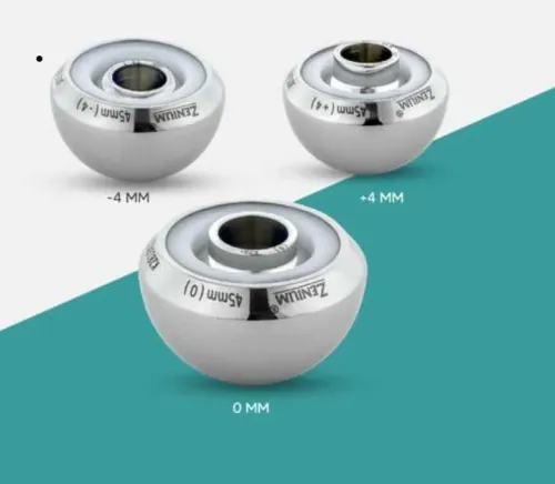
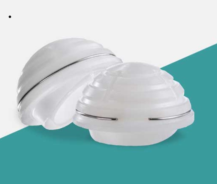 | Joint Replacement Acetabular Cup Cemented Manufacturer An Acetabular Cup Cemented Manufacturer produces acetabular components specifically designed for cemented fixation in total hip replacement... View product → |
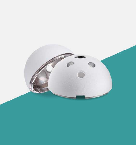 | Joint Replacement The Acetabular Cup is a key component in total hip replacement surgery, designed to fit into the hip socket (acetabulum). View product → |
Decide whether monitoring, urgent treatment or revision planning is appropriate
A stable radiographic finding with mild, unchanged symptoms and no serial migration belongs in planned review, not automatic revision. The treating orthopaedic team sets the interval for repeat examination and radiographs; an unchanged narrow radiolucent line does not carry the same urgency as documented movement.
Escalate to a specialist pathway promptly for:
- Progressive cup migration or changing inclination
- Cement mantle fracture or major osteolysis
- Recurrent dislocation or a periprosthetic fracture
- Wound drainage, fever, or suspected periprosthetic joint infection
Revision planning starts by defining the acetabular defect. Classify segmental and cavitary bone loss, retained cement, pelvic discontinuity, and the amount of host bone available for fixation. A loose cup with adequate bone stock is a different technical problem from a migrated cup with major osteolysis or pelvic discontinuity.
Give the revision team the complete implant card, operative report, serial radiographs, CT images, laboratory and aspiration results, culture history, and cement details. CT should answer a planning question such as defining bone loss or retained cement; metal artifact can limit interpretation, and CT does not replace infection testing.
When comparing replacement components, ask an acetabular cup manufacturer or distributor to verify the exact catalogue identity, liner compatibility, cement requirements, regulatory labelling, and traceability. Confirm material, lot or serial identifiers, cement brand and batch, mixing system, and any plugs or screws. Do not select by nominal size alone.
The reconstruction plan and verified compatibility must control procurement.
Frequently asked questions
What symptoms can indicate a cemented acetabular cup problem?
Groin pain, instability, dislocation, leg-length change and radiographic migration can indicate cemented cup complications.
Why are serial radiographs important before diagnosing a loose cup?
Sequential images show whether radiolucent lines, migration, wear or osteolysis are progressing rather than remaining stable.
How do clinicians determine which part of the hip implant is failing?
They combine symptoms, examination, serial radiographs, infection testing and implant records to assess the cup, cement mantle, bone and femoral component.
What does the cement mantle and operative record reveal?
The cement mantle can show fracture, defects or debonding, while the operative record identifies the cup, cement technique and components used.
When does a cemented cup problem require urgent treatment?
Urgent assessment is needed for suspected infection, acute dislocation, fracture, severe instability, neurovascular symptoms or rapid component migration.
Related product
 | Joint Replacement Acetabular Cup Liner Manufacturer An Acetabular Cup Liner Manufacturer specializes in producing liners that fit inside the acetabular cup during total hip replacement surgeries. View product → |