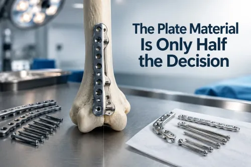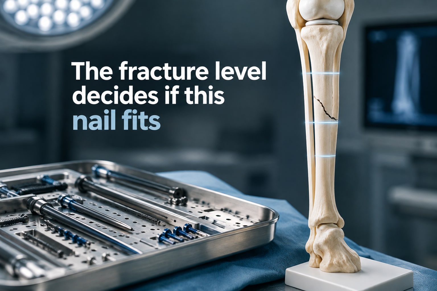
Choosing a tibial nail requires more than matching a fracture to a diameter and length on a catalogue page. You will be able to distinguish suitable fracture patterns from poor-fit anatomy, identify the measurements and soft-tissue conditions that change the decision, and compare nailing with plates, external fixation and staged treatment.
Key takeaways
- Confirm the nail’s hole geometry and sizes in the manufacturer’s instructions for use.
- Check fracture level, soft-tissue damage, infection risk and canal suitability before nailing.
- Measure tibial length, canal diameter and locking-hole position against the available nail sizes.
- Verify alignment, rotation, locking, imaging and implant records after fixation.
What a hyper interlocking tibia nail is designed to control
A hyper interlocking tibia nail is a locked intramedullary tibial nail designed to control fracture rotation, length and alignment through proximal and distal locking holes. “Hyper interlocking” is not a universal clinical standard, so confirm the nail’s exact geometry, hole locations and configuration in the manufacturer’s instructions for use.
Extra or strategically positioned holes can improve fixation in a short metaphyseal segment, where a shaft-focused pattern may leave too little bone.
Strong tibial fracture nail indications include:
- Unstable or displaced shaft fractures
- Comminuted or segmental fractures
- Short-oblique or long-oblique patterns
- Selected proximal-third or distal-third fractures
- Open fractures after appropriate debridement and treatment
- Fractures with bone loss
Static locking suits comminution, shortening risk and rotational instability because it fixes the nail to both main segments. Dynamic locking or later dynamization belongs to selected, relatively simple patterns; removing a screw does not correct a gap, malalignment or infection.
Blocking screws should be planned to narrow the effective canal and guide the nail through a metaphyseal fracture, not added as an afterthought. This is the mechanical basis of complex tibia fracture fixation. A stable, minimally displaced tibial fracture is not automatically an indication for surgery.
When fracture level and clinical condition make nailing a poor fit
A tibial nail is a poor fit when its locking points cannot control the fracture or when the patient’s condition makes definitive implantation unsafe.
A fracture entering the tibial plateau or ankle plafond needs independent anatomic reduction and fixation of its articular component; passing a shaft nail across the joint injury does not provide that control.
1. Treat a very proximal fracture cautiously. A short proximal segment risks procurvatum and valgus. Semi-extended or suprapatellar insertion, multiple proximal locking options and blocking (Poller) screws can improve alignment, but do not choose the implant from shaft-fracture specifications alone.
2. Assess a very distal fracture for enough bone to hold the planned screws. Confirm distal segment length, screw number and screw orientation, then check coronal and sagittal alignment. Limited distal purchase can make a nail unsuitable; blocking screws, fibular fixation or a plate may be necessary.
3. Review the interlocking nail clinical conditions before scheduling definitive fixation. Contaminated open wounds, severe soft-tissue damage, vascular or nerve injury, compartment syndrome, active infection, osteoporosis, pathological fracture and existing implants that obstruct the canal or nail path can alter timing or implant choice. These factors also qualify otherwise reasonable tibial fracture nail indications.
4. Use temporary external fixation when contamination is severe, infection is uncontrolled or damage-control stabilization is needed. Perform serial compartment examinations before and after fixation, even after inserting a nail.
A nail also does not guarantee early weight bearing in metaphyseal, comminuted or bone-loss injuries; progression depends on reduction, fixation purchase and soft-tissue condition.
Measurements that decide whether the listed nail sizes fit
A 260 mm nail is not automatically suitable for a short tibia, and an 11 mm nail is not automatically safe in a wide metaphysis. Measure the patient’s anatomy before treating the published 260–380 mm lengths and 8–11 mm diameters as options.
Obtain full-length anteroposterior and lateral tibia radiographs that include the knee and ankle. Add fracture-level CT when the fracture enters a joint or radiographs do not define the articular anatomy.
| Measurement | What it decides | Failure if ignored |
|---|---|---|
| Tibial length and proximal/distal segment length | Usable nail length and fixation on both sides | Joint penetration, prominence, or too few locking screws |
| Narrowest canal along the intended path | Diameter after considering shape, curvature, reaming strategy and stated tolerances | Nail impaction, cortical damage, or inadequate stability |
| Alignment, deformity and locking-hole position | Whether holes clear the fracture, existing hardware and usable cortical purchase | Malalignment, blocked insertion, or weak fixation |
For hyper interlocking nail tibia conditions, also check bone loss, unusual bowing, a wide metaphysis, previous surgery and skeletal immaturity against the device’s technical drawings and instructions for use. A planned blocking screw must narrow and guide the canal without obstructing the nail. Confirm every selected hole has enough bone for secure screw purchase.
How surgeons compare a hyper nail with other fixation strategies
The deciding question in complex tibia fracture fixation is not whether a hyper nail is newer, but whether its exact locking-hole pattern controls the available bone. Compare its manufacturer’s geometry and instructions for use with a standard locked intramedullary nail before treating “hyper” as a clinical indication.
| Option | Main advantage | Main limitation |
|---|---|---|
| Hyper interlocking nail | Load sharing with added locking choices for selected short segments | Short proximal or distal bone can still defeat alignment or screw purchase |
| Standard locked intramedullary nail | Less exposure of the fracture site and efficient shaft fixation | Metaphyseal or articular fragments may need independent control |
| Locking plate | Direct reduction and fixation of articular or metaphyseal fragments | Requires an approach through soft tissue that may already be damaged |
| External fixation | Minimal initial implant exposure and strong damage-control utility | Pin-site problems, frame management, and later conversion add procedures |
| Staged fixation | Allows irrigation, debridement, antibiotics, temporary stabilization, and soft-tissue coverage before definitive fixation | Delays final fixation and increases treatment stages |
A plate gains control where a nail struggles, especially at the joint, but its exposure can worsen a compromised envelope. External fixation fits severe contamination, uncontrolled infection, or damage-control needs; open fractures are not automatically excluded from nailing.
Compare alignment control, soft-tissue disruption, operative time, fluoroscopy exposure, weight-bearing progression, and possible implant removal or revision. Reamed nailing reduced reoperation versus unreamed nailing in a large trial of closed tibial shaft fractures; do not generalize that result to open fractures.
Early weight bearing follows a good reduction, adequate locking purchase, stable fracture pattern, and acceptable soft tissues—not the word “nail” alone. These factors remain central to tibial fracture nail indications.
What must be checked after fixation and before procurement
After fixation, confirm alignment, rotation, length, segment purchase and soft-tissue status on imaging and examination. Malalignment, delayed union, nonunion, nail bending or breakage, locking-screw breakage and loss of fixation become more likely with fracture gaps, severe comminution, infection, poor reduction, inadequate proximal or distal purchase and premature loading.
| Failure mode | Check | Consequence |
|---|---|---|
| Malalignment or fixation loss | Compare alignment and locking purchase with the operative plan | Revision may be needed |
| Delayed union or nonunion | Review serial radiographs, fracture gap and biological environment | Do not treat a gap by simply removing a screw |
| Nail or screw failure | Check for bending, breakage and progressive pain | Restrict loading and investigate stability and union |
Anterior knee pain remains a recognized late complaint. Accurate entry-point placement and avoiding proximal nail prominence matter more than any claim that a design eliminates pain.
Persistent drainage, deep infection, sequestrum or exposed hardware requires cultures, debridement, antibiotics and soft-tissue coverage; staged nail removal or exchange may be necessary. Changing locking screws alone does not treat infection.
Before a hospital or distributor evaluates a hyper interlocking tibia nail from Komal Health Care Pvt Ltd, request:
- Exact nail drawing and locking-hole map
- Indications, contraindications and surgical technique
- Sizing tolerances and compatible instruments
- Sterilization information and instructions for use
These documents define the interlocking nail clinical conditions and let buyers compare systems without selecting from diameter and length alone.
Related products
 | Bone Nails Our range of interlocking nail, intramedullary nail, fracture nail are can be used in 35 different cases of complex fractures. View product → |
 | Othopedic Implants Hyper Interlocking Nail For Tibia bone We ARe The manufacturer of Hyper Interlocking Tibia Nail For Bone, Size is in Diameter 8mm , 9mm , 10 mm , 11mm in length 260mm to 380mm Given... View product → |
Frequently asked questions
What does a hyper interlocking tibia nail control?
It uses proximal and distal locking holes to control tibial fracture rotation, length and alignment. Because “hyper interlocking” is not a universal clinical standard, verify the nail’s exact geometry in its instructions for use.
When can a tibial fracture be a poor fit for intramedullary nailing?
A nail may be a poor fit when the fracture level is difficult to control with the available locking configuration, the canal does not accept the listed sizes, or the patient has clinical problems such as severe soft-tissue damage or infection risk.
Which measurements determine whether a tibial nail fits?
Compare the tibial length, medullary canal diameter, fracture location, nail length, nail diameter and locking-hole positions. Use the manufacturer’s sizing chart and instructions for use rather than relying on the product name.
How do surgeons compare a hyper nail with other fixation strategies?
They compare control of alignment and rotation, fracture level, soft-tissue condition, bone quality, surgical access, available locking options and the risks of alternatives such as plates or external fixation.
What must be checked after fixation and before procurement?
After fixation, check radiographic alignment, rotation, length, screw placement and implant integrity. Before procurement, verify the exact model, diameter, length, hole configuration, instruments, sterile status, lot number and required documentation.